The 20-week scan is an exciting albeit nerve-wracking time for lots of people.
Most people know it as the one where you find out the sex of your baby (if you want to know, that is), but the most important reason for the scan is to find out whether there are any underlying issues with your baby that need further exploration. Hence why it’s sometimes referred to as an ‘anomaly’ scan.
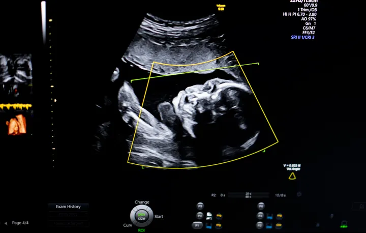
When you’re lying down with jelly on your tummy, watching what closely resembles a bean jumping about on the screen, it can seem like some kind of miracle that the sonographer can confidently determine the sex of your baby based on what they see – especially if you’re still trying to figure out what’s a leg and what’s the head.
We asked Kate Richardson, managing director and specialist sonographer at The Birth Company Harley Street and Alderley Edge, which is part of The Portland Hospital, to walk us through exactly how they do it.
She explains that before 12 weeks, it’s too early to notice any difference in sex by ultrasound. And although a sonographer may be able to identify the sex as early as 12 weeks, it’s not always a given and is very much dependent on how experienced they are, the quality of the ultrasound equipment, the position of the baby at the time of the scan and the mum’s build.
Between 12-14 weeks, the sonographer can obtain an ultrasound image in longitudinal cross section, and they can then assess something called ‘the foetal nub,’ which is basically the baby’s sex anatomy at this stage.
“For boys, the nub is positioned at an angle greater than 30 degrees, for girls the nub is positioned at an angle less than 30 degrees,” says Richardson.
“The quality and position of the image affect the likelihood of the correct sex being identified. It may be better to wait to find out when the result is more reliable, although it can be fun to compare your ultrasound images on nub theory websites and guess with your family.”
From 14 weeks onwards, your baby has developed more embryologically, explains the sonographer, so it is clearer to identify the difference.
“Baby girls continue to have a straight nub, which from the opposite angle can look like three white lines between the legs. Baby boys have now developed an appearance more consistent with a small scrotum and penis,” she says.
At 20 weeks, she suggests an experienced sonographer with good quality equipment should be able to correctly identify the sex of a baby by ultrasound in over 99% of cases. Although sometimes things don’t go as planned, for instance if the baby is in an awkward position.
At this stage, a baby girl’s anatomy remains as three white lines between the legs on an ultrasound, she explains, and the baby boy’s scrotum and penis also become more recognisable to the untrained eye.