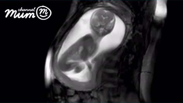
An incredible video is said to be the “next level of baby bump scans” using hi-tech MRI that is so detailed, it reaches right through the unborn baby’s body.
The video uses algorithms, magnetic fields and radio waves to create extra-high quality images of a tiny foetus. You can clearly see the baby - around 20 weeks old - fiddling with its umbilical cord, stretching its arms and turning its head.
It was captured by the iFIND project, a group of medical experts from around the world who made the world-first tech in a bid to improve antenatal scans.
“It can see the structures inside the body regardless of whether there’s bone, muscle or fat in the way and in some cases it can give us even more detailed images than ultrasound,” said Dr David Lloyd, clinical research fellow at King’s College London and part of the iFIND project.
“Importantly, it is also one of the few imaging techniques that is safe to use in pregnancy.”

The 24-second video scan ends with the unborn baby giving its mum a kick with both legs, causing her belly to wobble.
Unlike ultrasounds, the high-tech scan shows beneath the baby’s skin to get usable images from even the wriggliest babies.
Dr Lloyd added: “Taking pictures of a 20-week foetus while they’re still in the womb really isn’t that easy. For one thing, they’re very small.
“The foetal heart, for example, with all of its tiny chambers and valves, is only about 15mm long, so less than the size of penny.
“Ultrasound technology - used in all routine antenatal scans in the UK - is actually fairly good at visualising these tiny structures. It uses very high frequency sound waves which are reflected back (’echo’) from the structures inside the body to produce an image.
“In foetal ultrasound, the images produced can be excellent, but unfortunately that’s not true for every patient. Ultrasound has to be able to ‘see’ through the body to the parts of the baby we want to image, and that isn’t always easy.
“It will depend on the age of the baby, how they are lying in the womb, the size of the mother, and many other factors.
“MRI, which uses a strong magnetic field and radio waves to produce images, isn’t so limited.”

The team behind the new scan were given £10 million by the Wellcome Trust and the Engineering and Physical Sciences Research Council (EPSRC) to develop new scans.
It’s said to be the most “cutting-edge scan” available to date. Normally if a baby moved rapidly in a conventional scan - just like the baby in the vide above - you would get an unusable image and the mum might have to return for a re-scan.
Medics hope to develop the technology so it will eventually involve four probes which automatically move around the top of the mum’s tummy to get the best image.
This would mean more complications could be picked up earlier and even treated before the baby is even born.

The images, taken during a trial, were of an anonymous mum at a hospital in London. They were given to video parenting site ChannelMum.com to share with pregnant mums around the world.
Cathy Ranson, editor of Channel Mum said: “Scans are amazing as they help mums, dads and even other family members bond with their baby.
“There is nothing quite as emotional as seeing your unborn child moving inside you, and these MRI scans are taking images to the next level.
“They are truly breathtaking.”
The team behind the new scan technology are from Kings College London, St Thomas’ Hospital, Imperial College London, University of Firenze, Italy, the Hospital for Sick Children in Toronto and Philips Healthcare in Amsterdam.